Quick Overview:
Most teams building AI medical imaging software plan carefully for the part that turns out to be easiest. Training a detection model on chest X-rays is a solved engineering problem with mature tooling behind it. Proving that model works well enough for a regulator, and getting it to run inside a radiologist’s existing workflow, is where budgets and schedules actually go.
That gap is worth understanding before you write a line of code. Assembling a defensible dataset, establishing ground truth that survives scrutiny, and fitting your output into a PACS worklist without adding clicks are the parts that decide whether you ship.
This guide covers what building AI medical imaging software involves in 2026: the categories you can build, what the market for AI in medical imaging really looks like, the development process end to end, DICOM and PACS integration, the regulatory picture in both the US and EU, and honest numbers on cost and timeline.
AI in medical imaging means using machine learning models, usually deep learning and computer vision, to read or process medical scans. The software analyzes X-rays, CT, MRI, ultrasound, PET, or mammograms and flags findings, measures structures, or improves image quality before a clinician ever looks at it. In practice, AI in medical image analysis breaks down into three tasks: classification (is something there), detection (where is it), and segmentation (what is its exact shape and volume).
For anyone building software, though, the more useful question is not what the technology does. It is whether your software counts as a medical device.
That single distinction changes everything downstream. Software that reads a scan and suggests a finding is regulated as Software as a Medical Device (SaMD). Software that reconstructs an image, routes studies, or reduces noise often is not. Two products can use identical model architectures and face completely different regulatory burdens depending on what they claim to do.
So the first question worth answering is not “what model should we train.” It is “what exactly are we claiming this software does, and for whom?” Your intended use statement is a regulatory artifact, not marketing copy, and it constrains every technical decision that follows.
Four categories cover most of what gets built in AI in medical imaging and diagnostics. Each carries a different evidence burden, dataset requirement, and route to market.
This software scans incoming studies and pushes urgent cases to the top of the worklist. It does not diagnose. It says “look at this one first.”
Because it flags rather than concludes, the regulatory bar is lower and the path to market is usually the shortest of the four. Stroke and pulmonary embolism triage are the best-known examples. Ranking a queue so the urgent case surfaces first is also the least demanding form of AI in medical imaging to validate, since you are proving prioritization rather than diagnosis.
CADe marks suspicious regions. CADx goes further and characterizes them, for example classifying a lesion as likely benign or malignant.
This is the hardest category. You need large, diverse, expertly annotated datasets, and regulators will expect a reader study showing clinicians perform better with your software than without it. Mammography, lung nodule detection, and diabetic retinopathy screening all sit here.
These tools measure rather than interpret: cardiac ejection fraction, tumor volume, bone density, lesion tracking across prior studies.
Radiologists already do this work manually and inconsistently, which makes the value proposition easy to explain. The regulatory burden is real but usually lighter than diagnostic AI, because the software is automating a measurement rather than reaching a conclusion.
Denoising, super-resolution, low-dose CT reconstruction, and protocol optimization all fall here. The software never touches diagnosis, so many of these products are not regulated as devices at all.
If you want to enter medical imaging without a multi-year regulatory program, this is often the most practical entry point.
Here is something worth knowing before you put a market slide in your deck: analysts do not agree, and the gap is not small.
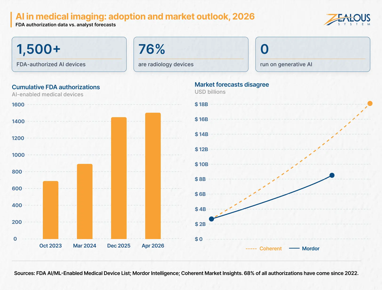
Mordor Intelligence puts the AI in medical imaging market at $2.16 billion in 2026, reaching $8.23 billion by 2031 at a 30.7% CAGR. Coherent Market Insights starts at a nearly identical $2.20 billion for 2026 but forecasts $17.77 billion by 2033. Same starting point, wildly different destinations.
Treat any single figure with caution, and expect a sharp investor to ask which report you used and why.
Adoption data tells a firmer story. The FDA has now authorized more than 1,500 AI-enabled medical devices, with 68% of those authorizations arriving since 2022. Radiology accounts for roughly 76% of the total, cardiovascular 10%, and neurology 4%.
One detail deserves more attention than it gets. As of March 2026, no FDA-authorized device uses generative AI or is powered by a large language model. Every cleared imaging product on the market today is a task-specific model. If your pitch rests on a foundation model reading scans, you are not late to a crowded market. You are ahead of the regulatory framework, and you should plan accordingly.
Demand pressure is genuine, too. Mordor projects the US radiologist shortfall will reach 19,500 by 2034, which is the structural reason hospitals keep buying despite thin outcomes evidence.
The measurable benefits of AI in medical imaging cluster in three places, and none of them is replacing a radiologist. Triage tools shorten time-to-treatment by surfacing urgent studies first. Quantification tools remove the variability that comes from different clinicians measuring the same structure by hand. And detection tools extend reading capacity in departments that cannot hire their way out of rising scan volumes.
Notice what is not on that list: outcome improvements. The evidence base for AI in medical imaging and diagnostics is still thinner than the marketing suggests, which is worth knowing before you build your clinical claims around it.
Not every imaging AI problem needs a new model. Three paths are worth weighing honestly before you commit engineering budget.
| Build Custom | License a Cleared Vendor | Integrate and Differentiate | |
|---|---|---|---|
| What You Own | The model and the clearance | Workflow and clinical UX only | Workflow, reporting, orchestration |
| Best When | No cleared equivalent exists, or the algorithm is your product | A cleared product already does the job | Imaging AI is one feature in a wider platform |
| Regulatory Load | Full submission, yours to run | Sits with the vendor | Minimal, unless you alter intended use |
| Time to Market | 12–24 months | Weeks to months | 3–6 months |
| Cost Profile | Highest upfront | Per-study or subscription fee | Moderate upfront, lower ongoing |
| Main Risk | Clearance timeline and evidence burden | Vendor dependency and margin squeeze | Thin differentiation if workflow is weak |
Building custom AI medical imaging software is the instinctive choice, and often the wrong one. Plenty of successful imaging companies are workflow companies with someone else’s model inside. If the choice is unclear, an AI consulting engagement that scopes the regulatory line first usually pays for itself.
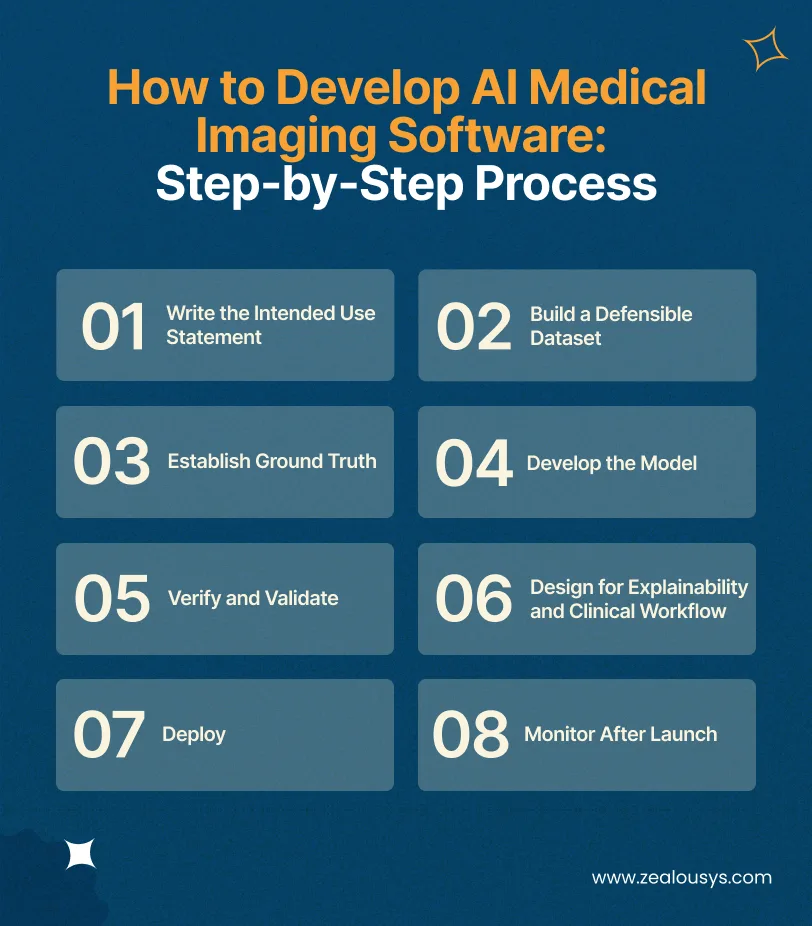
Most guides on how to build AI medical imaging software stop at the model. The sequence below reflects how imaging projects actually run, which differs from a standard AI software development process mainly in how much work sits before and after the model.
Define exactly what the software does, for which patient population, on which modality, and in what clinical setting. “Detects lung nodules on non-contrast chest CT in adults aged 18 and over” is a usable statement. “AI for radiology” is not.
Everything downstream, including your predicate search and your validation design, flows from this sentence. Write it first and revise it rarely.
You need thousands to tens of thousands of studies, and diversity matters more than raw volume. Data from a single hospital, a single scanner manufacturer, or a single demographic will produce a model that fails the moment it leaves that environment.
Source from multiple sites, across scanner vendors, and check demographic spread deliberately. De-identification has to be thorough, including burned-in pixel data in DICOM headers, which teams miss surprisingly often. Sound healthcare data governance practices here will save you months later.
Annotation is not data entry. Radiologists disagree with each other, and they disagree with themselves on re-read, so you need a documented protocol: how many readers per study, what qualifications they hold, and how disagreements get adjudicated.
Inter-reader variability is not a nuisance to eliminate. It is a property of the clinical problem, and regulators will want to see you measure it rather than hide it.
Task-specific convolutional networks still dominate cleared products, and frameworks like PyTorch, MONAI, and nnU-Net cover most imaging work. Transfer learning helps considerably when labeled data is scarce.
Federated learning is worth considering when hospitals will not release data at all: each site trains locally and shares only model updates. Framework choice matters less than validation discipline, though machine learning development experience in other regulated domains does shorten the learning curve.
Two different studies, often confused. Standalone performance measures your model against ground truth on a held-out, independent dataset. A clinical reader study measures whether clinicians perform better with your software than without it.
Diagnostic products usually need both. All of it happens under IEC 62304 documentation requirements, which is why V&V frequently costs more than model development.
Radiologists will not act on an unexplained flag. Grad-CAM heatmaps and similar techniques show which image regions drove the output, which lets a clinician cross-check rather than simply trust.
Design the interface around the existing reading workflow. Every extra click is a reason to stop using your product.
Cloud, on-premise, or hybrid, and the choice is often not yours. On-premise held a 63.2% share in 2025 because many hospital IT departments will not permit patient images to leave the building.
Build for both from the start. Retrofitting an on-premise deployment onto a cloud-only architecture is expensive.
Scanner upgrades, protocol changes, and shifting patient populations all cause model drift. Performance degrades quietly unless you are watching for it.
Log inputs and outputs, track performance against a monitored baseline, and define your retraining triggers in advance. This is not optional maintenance. It is a regulatory expectation.
This is where imaging projects lose their schedules. Your software has to receive studies, return results, and put those results somewhere a clinician will actually see them.
| Layer | Standard | What It Carries |
|---|---|---|
| Image Transfer | DICOM, DICOMweb | Studies in and out of your model |
| Storage and Retrieval | PACS, VNA | Where studies live and how you query them |
| Results into the Record | HL7 v2, FHIR | Findings pushed to RIS or EHR |
| Interoperability Rules | IHE Profiles | How the pieces are expected to behave together |
Four things reliably cause trouble.
Vendors populate optional tags differently, private tags appear without documentation, and two scanners in the same hospital can produce differently structured studies. Build tolerant parsing and validate against real clinical data early, because a pipeline tested only on public datasets will break on day one of a pilot.
A finding can return as a DICOM Secondary Capture, a Structured Report, or a message pushed into the radiology information system. Each choice changes what the radiologist sees and where they see it, which matters more to adoption than model accuracy does.
Triage products live or die on speed. A model that takes four minutes to process a stroke CT has missed the clinical window regardless of how accurate it is.
Many hospitals run installations that predate any AI integration standard, so you connect through a broker or middleware layer rather than directly. Both medical device software integration and integrating FHIR APIs cover those patterns in more detail.
Most imaging AI clears through 510(k), which requires showing substantial equivalence to an existing cleared device. De Novo applies when no predicate exists, and PMA covers the highest-risk products.
Worth understanding clearly: substantial equivalence is a comparator standard, not a clinical outcomes standard. A sponsor does not have to demonstrate that the tool improves patient outcomes. Clearance and clinical validation are separate things, and treating them as identical is a mistake both buyers and builders make.
A Predetermined Change Control Plan lets you specify in advance how your model may be modified after authorization. The FDA’s December 2024 guidance applies across 510(k), De Novo, and PMA.
Without one, retraining your model can mean a new submission. Write it before you submit, not after.
The European timeline moved, and a lot of published advice is now wrong. The Digital Omnibus, adopted by the Council on 29 June 2026, deferred the high-risk deadlines: standalone high-risk AI under Annex III shifts to 2 December 2027, while AI that is or sits inside an MDR- or IVDR-regulated device shifts to 2 August 2028.
Most imaging SaMD sits on the 2028 track, not the 2027 one, and most published guidance still gets that distinction wrong. Under Article 6(1), any AI medical device requiring a notified body, broadly MDR Class IIa and above, is automatically a high-risk AI system.
The good news is that the AI Act folds into your existing MDR conformity assessment rather than creating a second parallel process. MDCG 2025-6, published in June 2025, sets out how the two regimes interact.
ISO 13485 governs your quality management system, IEC 62304 your software lifecycle, and ISO 14971 your risk management. These are not paperwork you add at the end. Auditors will ask for records created during development, so the processes have to exist from the start.
In Europe, meeting these standards is what earns your CE marking. On the US side, HIPAA-compliant medical imaging AI requires encryption, access control, and audit logging on every system touching patient images. For broader context, our guide to medical device software development covers the wider compliance picture.
| Development Scope | Best For | Estimated Cost | Timeline |
|---|---|---|---|
| AI Proof of Concept | Validating a model on a limited dataset before committing | $20K–$40K | 2–3 months |
| AI Imaging MVP | Pilot deployment at one or two sites | $40K–$80K | 3–5 months |
| Production Platform | Multi-site rollout with PACS integration | $80K–$150K+ | 5–9 months |
| Enterprise Clinical Platform | High-volume, multi-modality, full compliance workflow | $150K+ | 9–15+ months |
What these figures do not include: These are software engineering costs. Annotation at clinical scale, a formal clinical reader study, regulatory consulting, and FDA submission preparation sit outside them, and you budget them separately. For diagnostic products, those regulatory costs can approach or exceed the development spend, and founders who miss this discover it in month three.
Our healthcare MVP development guide breaks down the pilot stage in more detail.
A working imaging AI team needs an ML engineer with medical imaging experience, a DICOM-literate backend engineer, a clinical advisor (usually a practicing radiologist), a regulatory lead, and QA with medical device software experience.
Those last two roles are the hardest to hire and the easiest to skip. Skipping them is also the most common reason submissions get delayed. Many teams cover the gap through healthcare IT staff augmentation rather than permanent hires.
The question that decides whether your product has a business, and it arrives late in most planning. Some imaging AI qualifies for CPT Category III codes, and a small number of products have secured New Technology Add-on Payment status in the US.
Most have neither, which means the hospital pays out of operating budget and you are selling against every other line item. Work out who signs the check before you build.
Narrow datasets: Models trained at one site with one scanner vendor fail elsewhere. Multi-site sourcing costs more upfront and far less than a failed validation.
Clearance mistaken for proof: A cleared competitor has not necessarily shown better patient outcomes. Understanding this shapes both your evidence strategy and how you sell against them.
Alert fatigue: A model with high sensitivity and mediocre specificity generates false flags until radiologists start ignoring it. Tune your operating point for the clinical workflow, not the leaderboard.
Model drift: Performance decays as scanners and protocols change. Without monitoring, you will not notice until someone else does.
Legacy PACS: Assume integration takes longer than your estimate. It usually does.
Four things separate teams that have shipped regulated imaging software from teams that have shipped software.
Predicate literacy: Can they read a predicate device summary and navigate the FDA’s cleared device database? A partner who cannot will not scope your submission correctly.
Production DICOM experience: Ask about specific edge cases, including private tags, burned-in PHI, and inconsistent vendor headers. Vague answers here are informative.
V&V documentation: Do they know what a submission requires? IEC 62304 records are created during development, not reconstructed afterwards.
Post-market support: A partner who disappears at launch leaves you owning the hardest ongoing obligation alone.
Also ask what a prospective partner does not do. Any team claiming to handle regulatory sign-off end to end is either misrepresenting the arrangement or has a partner they have not mentioned. Most of this comes down to whether the AI software development company you pick has built inside a regulated lifecycle before, or is about to learn on your budget.
Imaging AI rewards teams that plan for the parts nobody talks about. The model is tractable. Data, validation, integration, and post-market monitoring are the work.
At Zealous System, we build the engineering side of clinical AI products: model development, DICOM and PACS integration, and the software lifecycle documentation your regulatory team needs for a submission. Regulatory sign-off stays with your RA and QA function or a specialist partner, which is where it belongs. Our work spans healthcare software development and applied AI, including a pathology laboratory platform built for a diagnostic chain in India and an AI triage agent that cut manual review workload by 62% using the same queue-prioritization pattern imaging triage relies on.
If you are scoping an imaging AI build and want a realistic view of cost, timeline, and regulatory path before committing budget, that conversation is worth having early.
Between $20,000 for a proof of concept and $150,000+ for an enterprise clinical platform. You budget annotation at clinical scale, reader studies, and FDA submission preparation on top of those figures.
FDA review is typically 3 to 6 months for 510(k), but total elapsed time from development start to clearance is usually 12 to 24 months. Preparation, not review, takes the time.
It depends on your intended use. Software that detects, diagnoses, or triages is usually a medical device. Image enhancement, reconstruction, and workflow tools often are not.
Typically thousands to tens of thousands, though diversity matters more than volume. Data from multiple sites and scanner vendors beats a larger single-source dataset.
Public datasets are useful for development. Validation generally requires independent clinical data representative of your intended population. Synthetic data can supplement training but rarely substitutes for real validation data.
If it requires a notified body under MDR or IVDR, yes, as a high-risk system. Following the Digital Omnibus, most imaging SaMD must comply by 2 August 2028.
Build when the algorithm is your differentiation. Integrate when imaging AI is one feature within a broader platform and a cleared product already does the job.
Some products qualify under CPT Category III codes or NTAP in the US. Most do not, meaning the hospital funds it from the operating budget.
Our team is always eager to know what you are looking for. Drop them a Hi!

Pranjal Mehta is the Managing Director of Zealous System, a leading software solutions provider. Having 10+ years of experience and clientele across the globe, he is always curious to stay ahead in the market by inculcating latest technologies and trends in Zealous.


Comments